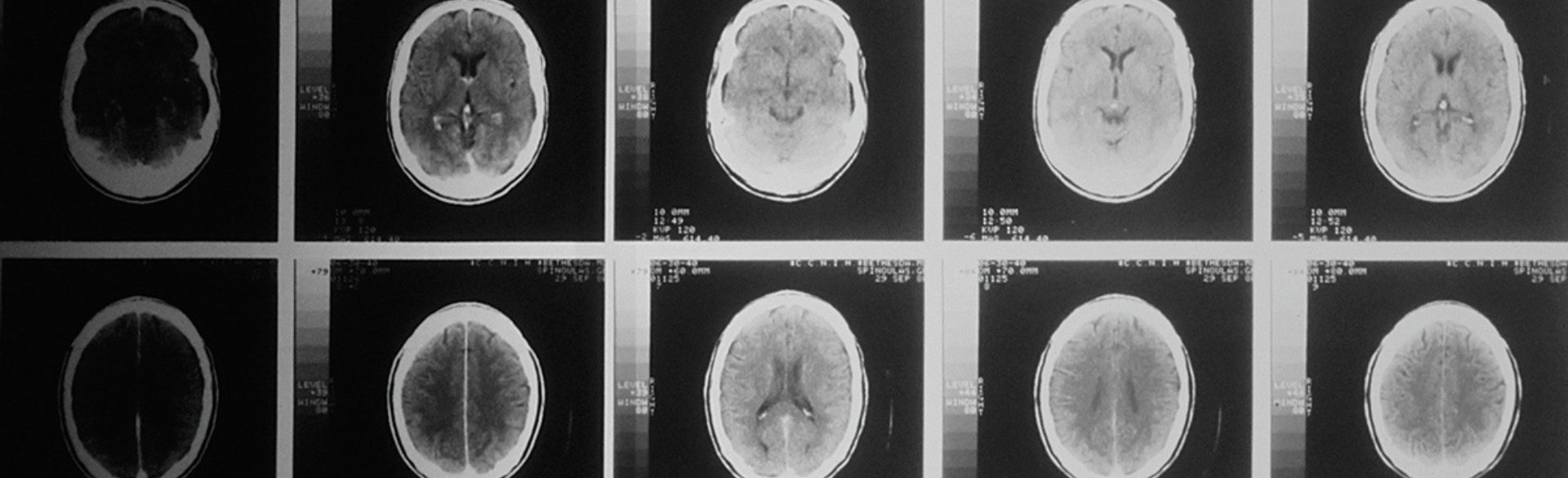
A new peer-reviewed paper presents the first data-driven sex-specific model of pediatric cranial bone development.
The findings reported today in PRS Global Open provide the first quantitative normative reference of local cranial bone development during the first ten years of life.
“Making this model publicly available to the scientific community is critically important for studying the phenotypes of cranial pathology and understanding how they may affect brain development in children,” said Antonio R. Porras, PhD, assistant professor in the department of biostatistics and informatics in the Colorado School of Public Health. Porras also works in the departments of pediatric plastic and reconstructive surgery and pediatric neurosurgery at Children’s Hospital Colorado. Both the school and hospital are located on the University of Colorado Anschutz Medical Campus.
Led by Porras, this study was funded by the National Institute of Dental and Craniofacial Research and performed in collaboration with Marius George Linguraru, DPhil, at Children’s National Hospital and Natasha Lepore, PhD, at Children’s Hospital Los Angeles.
Until today, there haven’t been any quantitative references that scientists or doctors could use to evaluate essential aspects of cranial development in children, such as local bone shape, thickness, mineral density changes, and the continuous processes of cranial suture fusion. These references are essential to study cranial suture anomalies such as craniosynostosis. They are also critical for evaluating and creating accurate diagnoses that can result in more personalized treatments.
“Previous studies on pediatric cranial pathology had been hindered by the lack of necessary quantitative normative references derived from large datasets. This had limited our understanding of the development of anomalies in the cranium and how they may affect the brain” Porras adds.
The paper proves the model’s accuracy predicting temporal development using an independent longitudinal dataset. The researchers also quantify developmental differences associated with sex and they study for the first-time suture fusion as a continuous process using objective data.
Porras concludes, “This new model will be essential to help us further understand cranial bone development in children and identify and quantify anomalies. It also constitutes the first necessary step to understand the coupled mechanisms of cranial and brain development.”
The entire paper is available here.
About the University of Colorado Anschutz Medical Campus
The University of Colorado Anschutz Medical Campus is a world-class medical destination at the forefront of transformative science, medicine, education and patient care. The campus encompasses the University of Colorado health professional schools, more than 60 centers and institutes, and two nationally ranked independent hospitals - UCHealth University of Colorado Hospital and Children's Hospital Colorado - that treat more than two million adult and pediatric patients each year. Innovative, interconnected and highly collaborative, the University of Colorado Anschutz Medical Campus delivers life-changing treatments, patient care and professional training and conducts world-renowned research fueled by over $650 million in research grants. For more information, visit www.cuanschutz.edu.