Can a lumpectomy patient’s chances of needing to return for a second surgery be reduced if the results of the operation are analyzed in real time, while the patient is still under anesthesia?
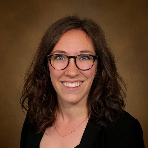
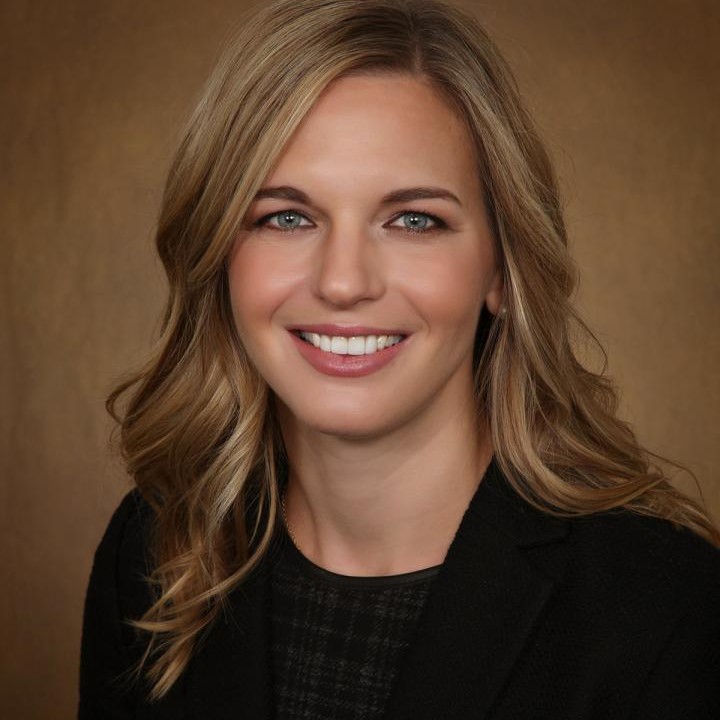
It’s a question that Sarah Tevis, MD, associate professor of surgical oncology in the University of Colorado Department of Surgery and CU Cancer Center member, has been working to answer for the past few years, since arriving at CU from another institution where the process — known as a multidisciplinary intraoperative margin assessment protocol —was the norm for lumpectomy patients with breast cancer.
“When we do a lumpectomy, there has to be no cancer at the edge of the lumpectomy specimen when we’re done,” Tevis explains. “The tissue we remove can’t have cancer at the edge, because if there’s cancer at the edge, then theoretically, there could be cancer remaining in the breast. We call that having clear margins.
“We need clear margins, but we don’t really know for sure until the pathology comes back after surgery, and that takes about a week,” she says. “I tell patients that 10% to 15% of the time, they’re going to have a positive margin, and we’re going to need to do a second surgery to remove more tissue. This process is asking, ‘Can we look more carefully at the margins during surgery to try to prevent that second surgery?’”
A new protocol paradigm
In a multidisciplinary intraoperative margin assessment protocol, the lumpectomy specimen is examined immediately after it is removed from the breast. An X-ray of the specimen is reviewed by a radiologist, while a pathologist slices the lump into sections to see if there is cancer remaining in any specific area that surgeons need to further excise.
“The radiologist looks at the X-ray of the slices, and the pathologist looks at the slices themselves, and then they call into the OR to tell the surgeon, ‘We think the margins are all clear,’ or, ‘We think you need to take tissue from this specific area, based on what we’re seeing,’” Tevis says.
Studying the timing
Madeline Higgins, MD, a resident in the CU Department of Surgery, is working with Tevis to study the feasibility of implementing multidisciplinary intraoperative margin assessment protocol. For recent research she presented at the 19th annual Academic Surgical Congress meeting in February in Washington, D.C., Higgins looked specifically at the timing of the process and how it compares to the traditional method of examining a specimen after surgery is complete.
“Ultimately we want to see how good we are at reducing rates of re-excision, which is the outcome we hope this is impacting, but right now we’re specifically looking at how long the process takes,” Higgins says. “I’m collecting time points — what time does the specimen get removed? What time does the X-ray get done? What time does the specimen get to pathology? If we can track each step of the process and how long it takes, we can find out if this process makes the surgery run longer than it should.”
That’s important, Higgins says, because surgeons don’t want patients to remain under anesthesia longer than they have to. So far, she has found that the real-time protocol extends most cases by around 15 minutes. It’s an acceptable trade-off for the benefits of the same-day examination.
“Ideally these patients won’t have to go back to the operating room because we’ve done more targeted selection of which margins to take out,” she says.
Getting more data
Higgins continues to gather information on the lumpectomy protocol and its timing, researching the feasibility of the process for a future publication.
“The next thing I want to do before I publish is talk to the people who have been part of the team about what processes have been challenging for them and what they feel has been good about the process,” she says. “It will help to define the weak points of this process and allow us to think about the recommendations we make to other surgery centers outside of the University of Colorado about how they can implement this protocol at their hospital.”
So far, Higgins says, the new process has generated excitement among the multidisciplinary team of surgeons, radiologists, and pathologists, who are excited at the potential it holds to improve care and help patients. In addition to the Academic Surgical Congress address in February, she plans to present her research at the American College of Surgeons’ Quality and Safety Conference in July.
“It’s been fun to work with this team of people who are excited about the work,” she says. “I didn’t know the pathologists or the radiologists very well before this, but we’re all taking care of breast cancer patients. It’s interesting to think about their care from these different angles and perspectives.”


.png)
