

You have probably done it: To avoid paying an extra bag-check fee for an upcoming flight, you try to fit everything into a small carry-on. You may opt to organize your suitcase strategically, so that your favorite outfits are easy to reach. If you simply throw everything in without thought, it’s a lot harder to find what you need upon arrival.
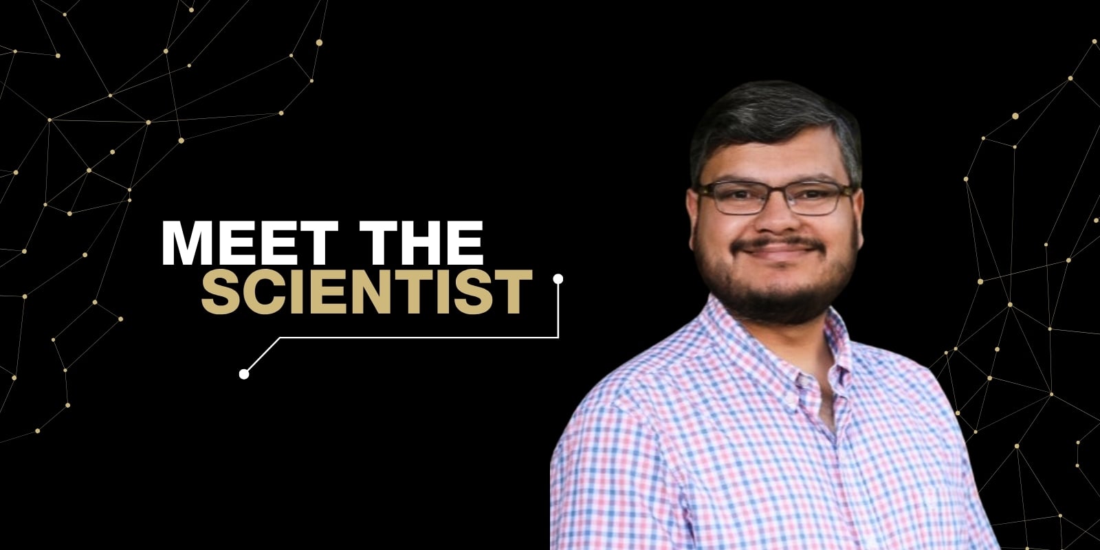
A research team led by Srinivas Ramachandran, PhD, associate professor in the Department of Biochemistry and Molecular Genetics at the CU Anschutz School of Medicine, is studying a similar problem: how the tremendous amount of genetic instructions for life, called DNA, are packaged into our very small cells.
Every cell in our bodies, from skin to stomach to lung cells, contains the same DNA. What makes each cell unique is which parts of the DNA are accessed and used by each cell type. Skin cells, for example, only need to interpret the DNA that codes for information needed to become and maintain skin cells.
This means that a large amount of DNA will go unused or never be accessed within each cell. This is where Ramachandran and his team come in. They aim to learn more about how the DNA is accessed before it’s used by the cell and if tiny marks or “snapshots” are left behind – basically flagging the sections of used DNA.
How do cells package 7 feet of DNA?
Each cell in our body individually contains nearly 7 feet of DNA, yet it must fit inside a nucleus that measures only a few micrometers across. In order for DNA to fit into the nucleus in an organized fashion, it must be very tightly and methodically packaged. To do this, DNA is wrapped around proteins called histones. Histones can be thought of as “beads on a string” (scientists have shown they really do look like individual beads on a long string). The string (DNA) is wrapped around the beads (the histones) as a way to package and organize the DNA.
In order for DNA to be utilized and turned into RNA, the “recipe card” for protein, transcription must occur. The transcription of DNA into RNA is a key step in all biological processes that require proteins. The first step is that DNA needs to be open and accessible to the cellular machinery that performs transcription. However, if the DNA is tightly packaged, the cellular machinery has a hard time reading the DNA to make RNA. It would be like finding your sweaters for winter that were stored in the drawer under piles of warm-weather clothes.
After it was discovered that cells package and organize their DNA in a distinct way, scientists then wanted to know how cells identify which DNA needs to be accessible to create the desired protein. Many scientists, including those in Ramachandran’s lab, approached this question in organisms and cells that were easy to work with, such as brewer’s yeast and cells taken from fruit flies.
To ask how DNA is packaged, they cut the DNA isolated from the yeast and fly cells with an enzyme that chewed up unpackaged DNA, leaving behind DNA wrapped around proteins. Using this DNA, they could make maps of which parts of the genome were open and which parts were closed.
Ramachandran’s team learned from working with fly cells that instead of completely unwrapping itself from its organized state, DNA can partially unwrap itself to become accessible to the transcription machinery. This partial unwrapping leaves behind a “snapshot” that can be used to further understand the cell and its potential disease state.
For example, if you had to quickly rummage through your summer clothes at the top of the drawer to reach your sweaters, you may leave behind a few “holes” where the winter clothes had been pulled out. Someone might walk in later and see you had a rushed morning trying to quickly dig out the winter clothes. This gives that person a “snapshot” of what happened earlier that day.
Using “snapshots” in DNA to understand disease states
What Ramachandran and his lab didn't know at the time was whether this same kind of snapshot, first seen in yeast and fly cells, would also show up in humans. It turned out it does.
When a cell dies, its DNA doesn’t just disappear. When the dead cells break down, their genomes are chewed up by enzymes, in a manner similar to how scientists were cutting genomes to study their packaging for decades. The result of this cutting is that the still-wrapped DNA can be found floating in the blood. These floating pieces of DNA are called cell-free DNA.
Ramachandran and his team use cell-free DNA as a snapshot of what the cell was doing right before it died. To analyze this cell-free DNA in the blood, they use a technique called cell-free DNA sequencing. This technique allows them to identify which cell type, and therefore which tissue, the DNA came from and offers clues about the disease state of the tissue. This approach allows scientists to study the potential disease state of tissues that lie deep within the body, without requiring invasive surgery to obtain a biopsy. This method can be thought of as a “liquid biopsy” because it uses blood samples instead of tissue samples.
How can a ‘liquid biopsy’ lead to tailored treatment?
Imagine someone sitting in an oncologist’s office, waiting to learn not just whether they have cancer, but which kind, because that answer determines which treatment gives them the best chance to overcome it. What the oncologists need is a molecular snapshot of the cancer, so they can choose the right treatment to attack it. One such molecular marker in breast cancer is estrogen receptor alpha, or ER. By knowing which form of breast cancer the individual has – ER+ or ER- – it allows their healthcare team to better tailor treatment.
Work done by Ramachandran and his lab has shown how a “liquid biopsy” could potentially be used to understand the class and severity of ER+ breast cancer. By analyzing the cell-free DNA found in the blood of individuals with breast cancer, more information can be gathered about their ER status and allow scientists to understand the speed of tumor development and classify tumor subtypes. One day, this may allow for earlier cancer detection and more personalized treatment.
What’s next for the Ramachandran lab?
Ramachandran and his lab are expanding this idea of a “liquid biopsy” into other fields of biology. They are currently working to understand how this technique could be used to understand the aging process of various tissues. For example, you may feel and appear healthy, but this technique may be able to identify early signs of an aging heart. By identifying heart problems early, you can intervene with lifestyle changes or other therapeutics to potentially slow, or even reverse, these signs of aging.
Scientists are working hard to identify interventions that slow the effects of aging, but these studies take decades to tell if the interventions are beneficial. Ramachandran’s lab has a goal to develop non-invasive blood tests that could tell in a matter of months if a lifestyle change or a new supplement is actually improving your health.
If you want to learn more about the scientist, check out his official CU Webpage.
To learn more about the Ramachandran Research Lab, check out the website here: Ramachandran Lab.


.png)