
Prostate cancer is the second most common and deadly cancer in the United States. The American Cancer Society estimates that 1 in 8 men will receive a prostate cancer diagnosis in their lifetime. Treatment techniques range from aggressive therapies such as radical prostatectomy or radiation therapy to targeted therapies that treat only the affected cancer cells.
The PSMA/PET Scan is a newly acquired imaging technique at the University of Colorado Cancer Center that provides a more accurate picture of what patients are dealing with. It was recently approved by the Food and Drug Administration (FDA), and it has proven to be an important tool for accurately detecting prostate cancer.
The ability to locate the affected regions with this precision significantly impacts the treatment team's ability to individualize treatments. This is especially true for determining if cancer has metastasized or moved to other areas outside of the prostate. Targeted therapies are less invasive and can significantly reduce common adverse side effects. Localized treatments isolate the cancerous cells from healthy tissue. This reduces the exposure and risk of developing negative side effects.
Paul Maroni, MD, University of Colorado Cancer Center member and associate professor of urology at the CU School of Medicine, explains, “This imaging technique is useful in many instances when treating men with prostate cancer. First, it’s a better staging study for men diagnosed with high-risk diseases. It’s also a great study for men with rising PSA levels following treatment with radiation or surgery to see where cancer might be.” He says, “We have been studying and watching the progress of the PSMA PET technology for years and are very excited to be able to offer this to our patients.”
How does it work?
Bennett Chin, MD, professor of radiology and nuclear medicine at the CU School of Medicine, works with the multidisciplinary team at the CU Cancer Center on these imaging techniques. He verifies how the Prostate Specific Membrane Antigen (PSMA) positron emission tomography (PET) works by administering a radioactive tracer drug (68Ga-PSMA or 18F-PSMA) to patients about an hour before the PET imaging scan. The drug works by attaching to the PSMA antigen that is overexpressed in prostate cancer cells. The tracers bound to the cancer are now seen on imaging during the PET scan. The PSMA/PET scan drug is manufactured here on the CU Anschutz Campus. This technique has improved the standard of care for the multi-disciplinary team treating patients at the CU Cancer Center.
Further studies are underway for researchers working at the CU School of Medicine that look at the benefits of combined PET and MRI. Our campus houses a very unique, simultaneous system that uses magnetic resonance imaging (MRI) to layer multiple image types, providing another set of valuable images to improve cancer detection and localization (PET/MRI). We have already started to use the PSMA PET/MRI technique on select patients.
How does the PSMA/PET differ from other imaging?
From the patient perspective, no additional side effects or treatments are associated with receiving this new technology. Maroni explains, “This provides extra information that stages their cancer more accurately. It’s not any quicker. More accurate? Very.”
“This provides extra information that stages their cancer more accurately.
It’s not any quicker. More accurate? Very.”
The standard imaging technique used to test for metastatic prostate cancer includes a diverse list of diagnostic testing that includes:
- Bone Scans – Prostate Cancer can spread to areas outside the prostate gland. Advanced cancer can spread to the bones.
- Computed Tomography (CT) Scans – Contrast injection follows a form of X-ray to provide a cross-section of the pelvic region.
- Magnetic Resonance Imaging (MRI) – Utilizes using powerful magnets to scan soft tissue and tumors for internal imaging.
- Positron Emission Tomography (PET) scans – Injection of a radioactive tracer followed by scanning procedure.
- Transperineal MRI Fusion Biopsy – Combines ultrasound and MRI scans.
Compared to these techniques, the PSMA/PET has shown to be more effective at isolating tumors and affected cells throughout the entire pelvic region. These scans can detect cancer in bones and soft tissue and can identify small lesions in and around the prostate gland. It aids in monitoring advanced cancer and identifying metastases due to its ability to pinpoint cancer missed by standard imaging. Thus, replacing the potential for surgical biopsies. Studies indicate that it is helpful in the early detection of recurrence in men who previously required radical treatments.
The images below demonstrate how the PSMA/PET more clearly identifies the location of prostate cancer.
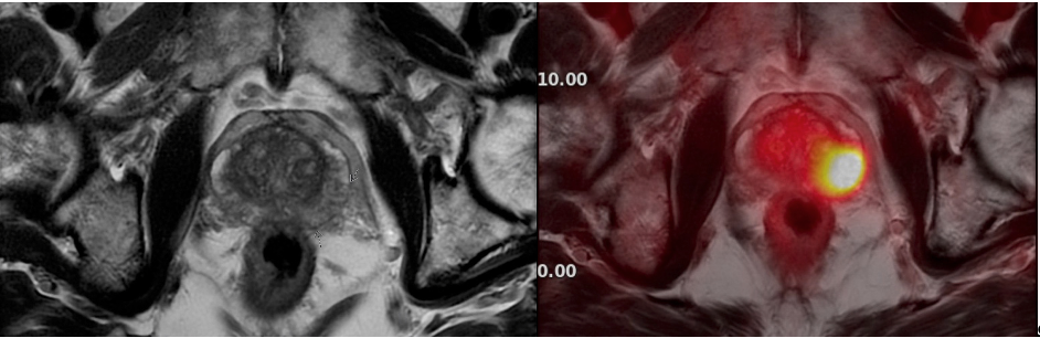
(Left): MRI only. (Right) PSMA/PET MRI fused image clearly shows prostate cancer.
With the PSMA/PET imaging available at the University of Colorado, the community now has access to the leading technology in prostate cancer imaging. The range of benefits includes less invasive treatments, the ability to locate affected areas more accurately, and, ultimately, the ability to determine the best course of treatment.
“The PSMA/PET and hybrid imaging techniques, such as the PET-MRI, are the future of how we will be doing prostate cancer imaging. There are a lot of exciting opportunities now concerning targeting treatments.”“The PSMA/PET and hybrid imaging techniques, such as the PET/MRI, are the future of how we will be doing prostate cancer imaging. There are a lot of exciting opportunities now concerning targeting treatments.” Maroni concludes, “Patients can look forward to receiving specific-individual treatments that will improve outcomes.”