University of Colorado Cancer Center leader Daniel LaBarbera, PhD, has received an innovation pilot grant from the cancer center’s Shared Resources program to develop complex 3D tissue and organoid models for high-throughput drug discovery and high-content imaging.
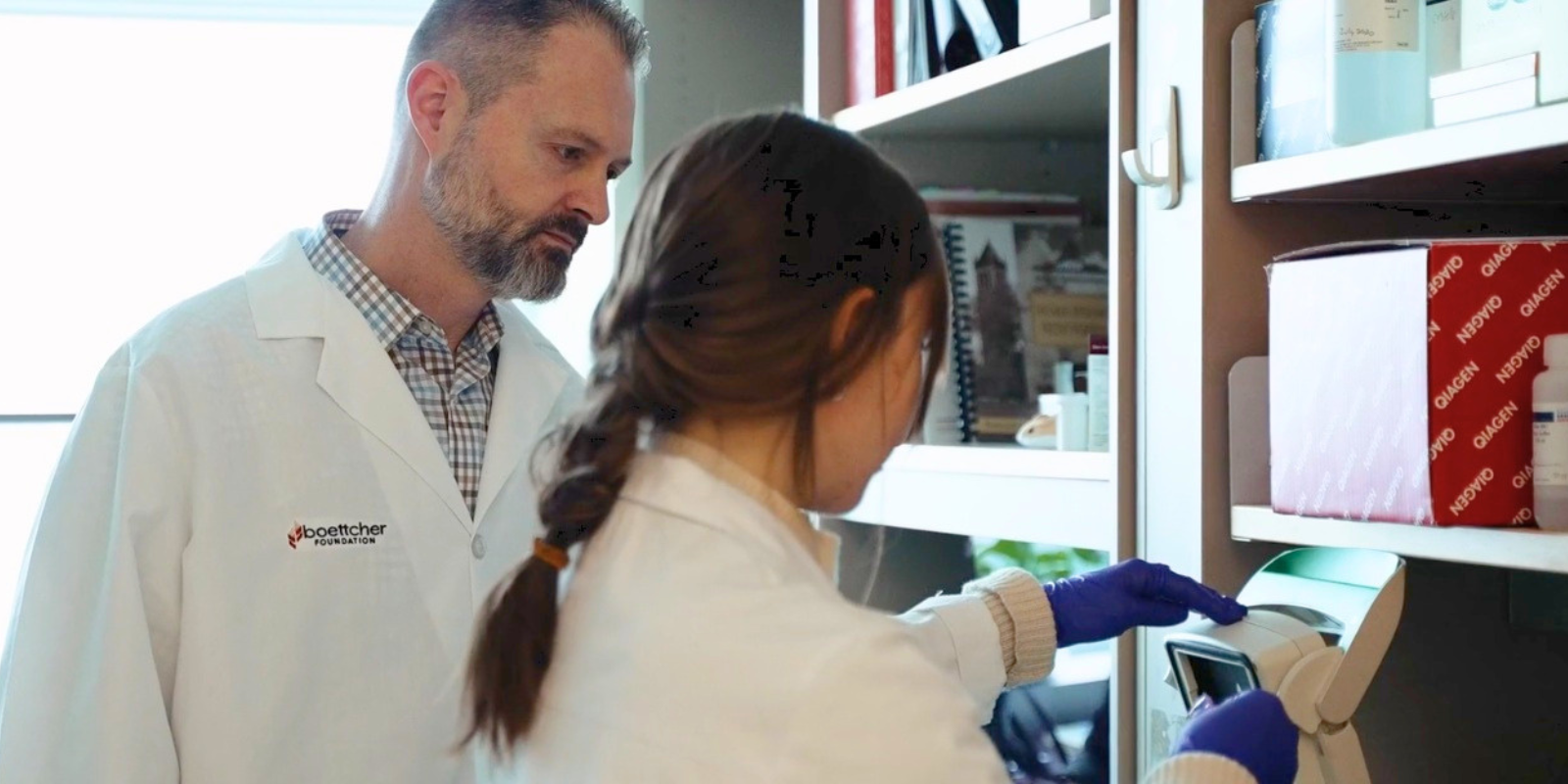
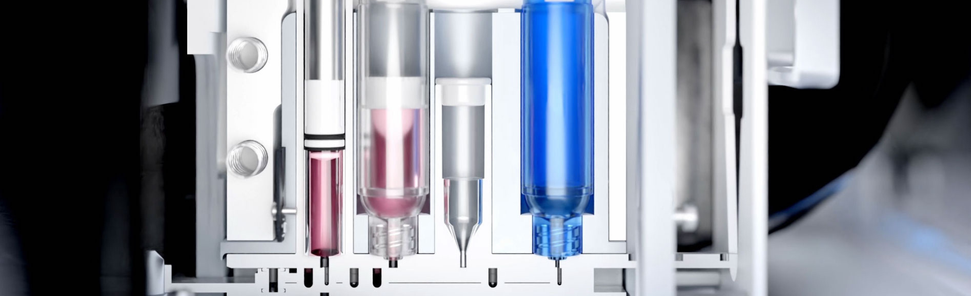
LaBarbera, a professor in the Skaggs School of Pharmacy and Pharmaceutical Sciences and director of the Center for Drug Discovery on the CU Anschutz Medical Campus, is director of the Drug Discovery and Development shared resource at the CU Cancer Center. LaBarbera will work withHector Esquer, PhD, an instructor in the Center for Drug Discovery and Drug Discovery and Development shared resource, to install and operate a 3D bioprinting instrument to create a variety of complex 3D models of tissue and tumor organoids with funds from the innovation pilot grant. Created by CellInk, the bioprinter automatically and precisely dispenses a mixture of cell suspensions and extracellular matrices and other “bioinks” into well plate formats.
“Within this year, we hope to be the first academic center in the country to have an instrument like this integrated with a robot to do cell tissue printing in high-throughput screening plates,” LaBarbera says. “We’re going to use that to develop three-dimensional culture models that mimic some aspect of tumor biology, including metastasis or invasive potential.”
Cancer in three dimensions
The new technology, he says, will offer advantages over the in vitro techniques currently used by many cancer center researchers.
"One of the big areas for cancer biology is utilizing patient samples to develop patient-derived organoids,” LaBarbera says. “The problem is that cancer isn't flat. Tumors don't grow in a monolayer; they grow in three dimensions. This will allow us to combine tumor cells in a three-dimensional matrix that mimics the microenvironment of the tumor, but we can also add other cells, like immune cells, fibroblasts, and other types of cells that are common in the microenvironment that promote tumor progression.”
Rapid drug testing
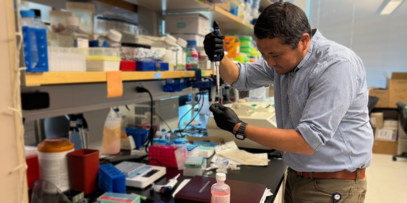
The robot-operated bioprinter also offers researchers the opportunity to quickly test multiple drug compounds against different cancer types to see which is most effective.
“We work in microplates, which typically have 384 wells,” LaBarbera says. “In each well, you could have a tumor organoid, or multiple tumor organoids, of different complexities. In house, we have 125,000 drug-like compounds. So we can screen a single potential drug candidate to see which drugs are most effective against the biological activity of that tumor. What's really cool with the tissue printer is we can print them precisely on the plate, so we know exactly where they are, and we can go back to them and image through them in a very rapid manner.”
The Drug Discovery and Development shared resource already was leading the way among academic centers when it came to custom automation and high-throughput drug discovery, LaBarbera says. The CellInk technology will put the CU Cancer Center even farther ahead.
“For some period of time, we will have a unique resource for investigators here in our cancer center, that no other academic institution is going to have,” he says.