Behind much of the groundbreaking research performed by members of the University of Colorado Cancer Center is the cancer center’s Shared Resources program, an assemblage of 12 facilities that offer researchers access to technologies and expertise that too complex and too expensive for an individual lab. Available to all CU Cancer Center members, as well as community researchers, on a fee-for-service basis, the Shared Resources include programs in oncologic imaging, biostatistics and bioinformatics, flow cytometry, mass spectrometry, and more.
Strength in equipment
Increasing the effectiveness of the Shared Resource programs is an array of cutting-edge equipment — everything from biomedical imagers to cryo-electron microscopes and mass spectrometers. Under the leadership of Associate Director Natalie Serkova, PhD, the Shared Resources have had extraordinary success over the past five years securing National Institutes of Health (NIH) shared instrumentation S10 and R24 awards — grants specifically designed to support the purchase of state-of-the-art, commercially available instruments to enhance the research of NIH-funded investigators.
“Over the past five years, we have received five NIH instrumentation grants ranging in price from $250,000 to $2 million,” Serkova says. “We have initiated a workshop on how to write S10 grants, because they are completely different from any other NIH grants. The average success rate for S10 grants is around 18% nationwide; with our S10 writing program, 100% of our applications have been funded."
Among the ingredients of a successful S10 application, Serkova says, are a strong group of Shared Resources researchers with the technical expertise to operate the equipment, solid scientific need and benefit of the requested instrumentation to NIH-funded researchers, and strong institutional commitment to Shared Resources.
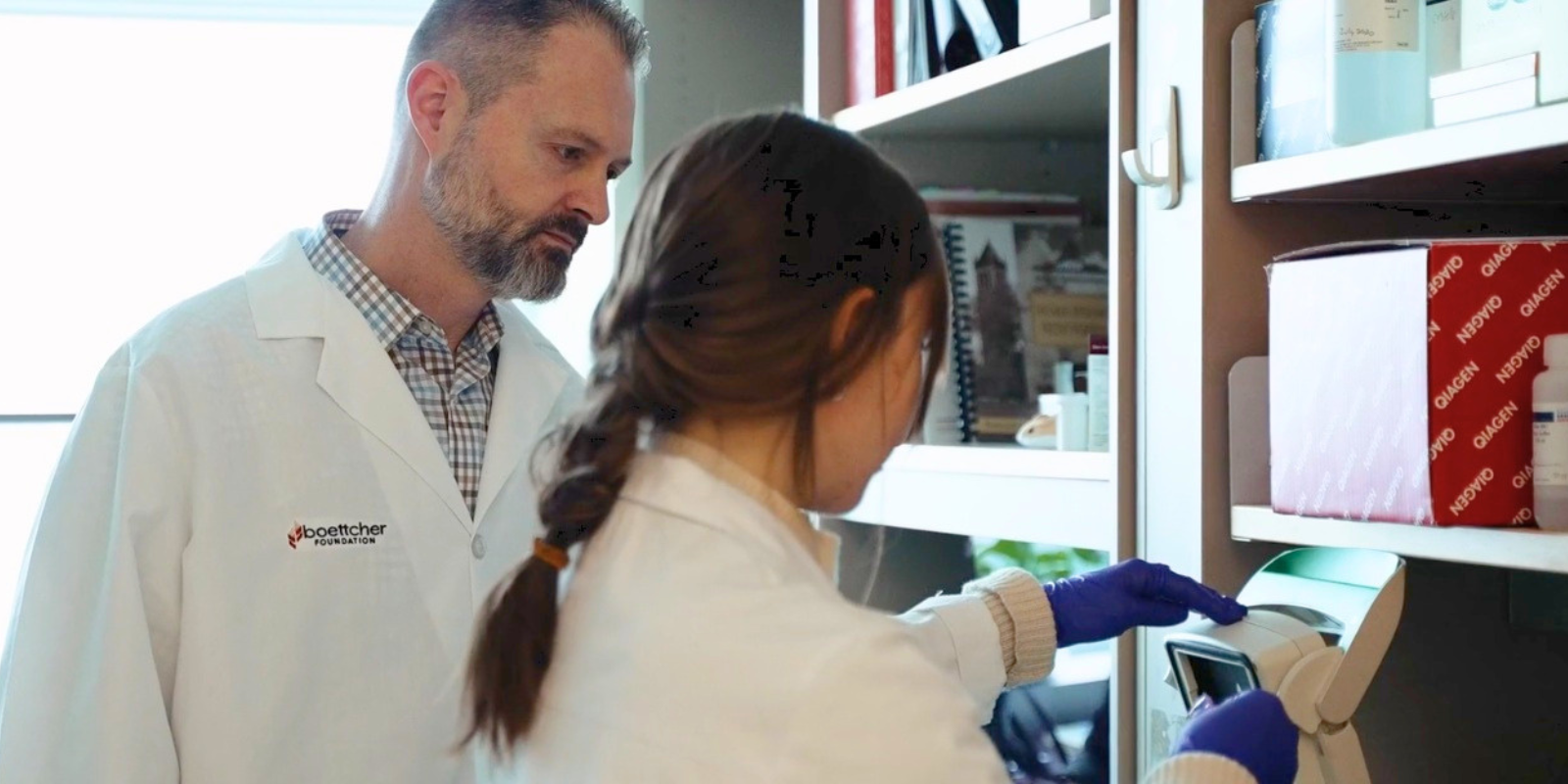
 Natalie Serkova, PhD, and an assistant work on a PET/MR hybrid scanner in the Animal Imaging Shared Resource.
“Researchers are not always familiar with technology,” she says. “Sometimes we have to explain the technology and its advantages that will help them answer their specific cancer biology or genomics question.”
New arrivals
The two most recent equipment acquisitions in Shared Resources are a Talos Arctica cryogenic transmission electron microscope, which yields 3D structures of large macromolecules, by the Structural Biology Shared Resource, and a Lunaphore COMET, a fully automated sequential multiplex instrument for staining, imaging, and image analysis of cancer and immune cells, by the Human Immune Monitoring Shared Resource (HIMSR).
“The COMET’s primary application is the study of tissue microenvironments,” says Kimberly Jordan, PhD, HIMSR Associate Director. “We're looking at what type of cells infiltrate tissues and the molecules those cells are producing — their activation state or activity level, and also where they're located spatially — so we can analyze not only what makes up the tissue microenvironment, but how they're organized relative to other cells and structures within the tissue.”
New view of the microenvironment
Jordan and her fellow HIMSR Director, Jill Slansky, PhD, recently received a CU Cancer Center Shared Resources Innovation Grant to develop antibody panels that will be used on the COMET to study cancer microenvironments. Jordan and Slansky will work with CU Cancer Center members Virginia Borges, MD, and Traci Lyons, PhD, on a breast cancer panel and with CU Cancer Center member Benjamin Bitler, PhD, on an ovarian cancer panel.
“For the breast cancer cases, we'll be comparing expression levels of different proteins that the group thinks are indicative of outcomes and then how those relate to hormone expression and other tumor marker expression, as well as the organization of the immune cells that surround and infiltrate next to the tumor cells,” Jordan says.
Similarly, Bitler says, his team will use the COMET to examine the ovarian cancer tumor microenvironment to look for ways to help patients who are resistant to traditional therapies.
“Therapy-resistant ovarian cancer is a major clinical challenge,” Bitler says. “Even within the same tumor, multiple mechanisms can contribute to therapy resistance. The Lunaphore COMET technology provides a powerful approach to identifying these different mechanisms within a single tumor. We are confident that the data from COMET will help identify novel combination therapies that could overcome therapy resistance and improve patient outcomes.”