
“Our goal is to develop robust and efficient software to reduce human suffering.”
— Greg Way, PhD
Before a pharmaceutical treatment or genetic intervention makes its way into the healthcare system, it has been rigorously tested in model organisms and clinical trials.
Yet, in deciding which treatments even reach this stage, researchers are faced with a major challenge: in a theoretically infinite pool of potential treatments, how can researchers effectively select the best candidates for further research and testing?
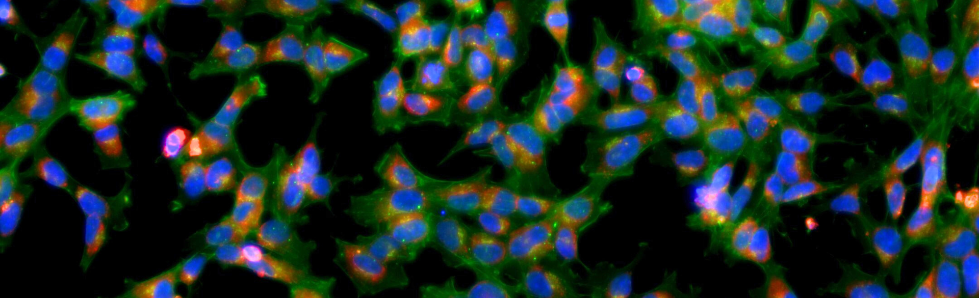
One approach is image-based profiling, a method to study the morphology—the shape and structure—of cells. In many diseases, cells undergo changes in shape and structure, causing them to display differently than a healthy cell. These phenotypic, or observable, changes can be seen by microscope imaging.
At the Department of Biomedical Informatics (DBMI) at the University of Colorado Anschutz (CU Anschutz), researchers are leveraging image-based profiling to efficiently test thousands of compounds, narrowing down the pool to find the best candidates for future research.
Cell Phenotype: The observable set of characteristics of a cell, including its physical, functional, and biochemical properties. Cell Morphology is the structural characteristics of a cell, including the shape, structure, form, color, size and orientation of the cell and its organelles. These characteristics can be visualized and measured using microscopic images, which can suggest underlying biological patterns and processes.
Led by Gregory Way, PhD, MS, associate professor of biomedical informatics at CU Anschutz, the Way Lab has developed a suite of open-source tools that support the image-based profiling pipeline. These tools allow researchers to analyze large-scale databases to define specific phenotypes, or observable changes, associated with disease. Researchers then evaluate how potential therapies such as genetic- or drug-based perturbations might shift the cell back towards a healthy phenotype. This helps them to uncover new therapeutics for diseases with unmet need.
This is the eighth installment in our ongoing series spotlighting the DBMI Wall of Software, an interactive online hub showcasing the latest open-source software and data tools for researchers. View the Wall of Software here.
Read on to learn more about image-based profiling, uncover the unique way that the Way Lab approaches open-source scientific software development in the sciences and explore some of the foundational tools in their image-based profiling platform.
A Brief Summary of Image-Based Profiling
Image-based profiling is a powerful method to evaluate and quantify cell phenotypes. Image-based profiling begins in a wet lab, where researchers leverage complex microscopic camera machines to automatically capture images of cells across hundreds of wells—sometimes across hundreds of plates.


Researchers in the labs can then apply different compounds or interventions across the wells, allowing them to efficiently take images that can then be used to evaluate the effects of various interventions on the cells.
After these images are taken, they are put through a feature-extraction software. This software will segment out individual cells, then it will calculate the morphological traits of the cells at a single-cell level. This data comes in large, tabular datasets that can then be analyzed for drug-discovery.
Open-Source Software Development in the Sciences: The Cytomining Ecosystem
The Way Lab develops open-source software, which means that the software’s codebase is transparent and visible to the public, it is free to use and reuse and it has the opportunity to be collaboratively developed by other developers in the community.
“The tooling that we build—the open-source software—is important for us because we use it every day. But we also want the whole community to benefit from our open-source tooling,” emphasized Way.
A Unique Approach to Open-Source Software
All of the Way Lab’s software tools contribute to the overall mission of image-based profiling, making every tool a component of what they call the ‘Cytomining Ecosystem.’
Way explained that “although all of these software tools contribute to the same mission of image-based profiling and phenotypic drug discovery, we have built a system with multiple tools because each tool is complex enough to justify being isolated for functional and maintenance reasons. Every tool has a core developer who is actively maintaining the project and making useful enhancements to tool functions.”
Distributing the tools in this manner ensures that each tool receives plentiful attention and support, and also allows the outputs of each tool to be used across varying contexts. Additionally, distributing the tools across individual developers facilitates collaboration across the team. Because the inputs and outputs of these tools are in conversation with one another, it is essential that the team works collaboratively to ensure the Cytomining Ecosystem creates a fluid, cohesive pipeline to support image-based profiling.
Foundational Tools in the Cytomining Ecosystem
The Way Lab has developed a variety of tools as part of the Cytomining Ecosystem. Here, we will explore a few of the core tools that build a powerful foundation for image-based profiling and phenotypic drug discovery, including Cytotable, coSMicQC, Pycytominer and Buscar.
CytoTable
CytoTable is a Python package that helps to prepare data for analysis by harmonizing the outputs of images and image-features into a consistent format. This process allows researchers to spend less time wrangling data, and more time focused on biological discoveries.
Dave Bunten, software engineer at DBMI, leads the development and maintenance of CytoTable.
Bunten emphasized that one innovation that CytoTable offers is the ability to output data in either anndata or Parquet files.
Parquet files were one of the first formats the Way Lab introduced to CytoTable, because Parquet files are fast and consistent.
“When we’re dealing with a lot of data like this, we need any data that we access later to be high performance. Parquet is special because it’s a columnar table type—which is different than, say, a spreadsheet file, in that you can access individual columns without having to open up the full contents of the table,” explained Bunten. This innovation allows the data to be accessed quickly, without overloading the resources of the computer being used.
“Parquet is also high performance in the sense that data are always the same type,” said Bunten. “Everytime you open a Parquet file, the data that’s kept inside that file is read in the exact same way into memory... These datatypes give you consistency across multiple different programming languages, which means that no matter whether you’re writing in Python, R, Julia, Rust—you always get the same data out of that same file. This means that for scientific work, we get consistency and reproducibility, which is a major advantage that we have through CytoTable.”
After data is prepared with CytoTable, it can then be leveraged by coSMicQC and Pycytominer to continue with the image-based profiling workflow.
CoSMicQC
CoSMicQC is a Python package for performing quality control for single-cell morphology profiles. After data is prepared in CytoTable, researchers can leverage coSMicQC to catch common problems and remove low-quality data from datasets before downstream analysis.
Jenna Tomkinson, senior quantitative cell biologist in the Way Lab at DBMI, leads the development and maintenance of coSMicQC, which helps researchers evaluate cell segmentation profiles.
Cell segmentation is the process of identifying individual cell boundaries from microscopy images. “A good example of segmentation is how people, with their smartphones, can actually segment things and make stickers,” explained Tomkinson. “They just take a photo, and click and hold the photo, and it will segment and outline it.”

“In our case, it’s not as simple as this, because we need to do this across hundreds of cells in one image. So, we need more complex and sophisticated algorithms,” said Tomkinson.
Within cell segmentation, technical deviations, artifacts or erroneous segments can arise, which can interfere with analysis. These issues can stem from many sources, including microscope issues and contamination from dust or unwanted material.
“CoSMicQC helps label or remove incorrect segmentations before downstream analysis, which supports cleaner, more accurate results,” explained Tomkinson.
After data has been processed through coSMicQC, it is then ready to be processed through Pycytominer.
Pycytominer
Pycytominer performs the image-based profiling bioinformatics pipeline; it is the core Python package in the Cytomining ecosystem. Pycytominer takes outputs from CytoTable and CoSMicQC and other experimental metadata to process single-cell features, getting them ready for downstream analysis. Way leads the development and maintenance of Pycytominer.
“Pycytominer is going to take in features that we’ve already extracted from microscopy images and process them through an image-based profiling pipeline,” explained Way. “That means it’s going to annotate the single cell: it’s going to tell which treatment and which well the cell came from, it’s going to normalize the single cells (to make sure that all of the features are on the same scale), and then it’s going to perform feature selection—which makes sure all of the features are informative in the profile.”
One unique feature of Pycytominer is its ability to perform batch effect correction. “Batch effects are technical artifacts that might arise from technical differences across different plates and different batches. Pycytominer has algorithms that perform batch effect correction.”
Pycytominer outputs a series of Parquet files that can be analyzed downstream.
Buscar
Buscar, or (Bioactive Unbiased Single-cell Compound Assessment and Ranking), is a Python package that helps researchers identify biologically active perturbations. Buscar is also a play on words on the Spanish and Portuguese verb “buscar,” which translates as “to search” or “to look for.” Buscar helps researchers search for and prioritize effective compounds in large-scale drug screening experiments. In these drug-screening experiments, researchers apply thousands of potential therapeutics to diseased cells. Buscar helps researchers identify which therapies are the most promising to make diseased cells look healthy again.
Erik Serrano, PhD, researcher at DBMI, leads the development and maintenance of Buscar.
Buscar leverages a unique approach to find drug candidates for a specific disease. “We generally use summary statistics to compare and ask: ‘is this drug working, or not?’” said Serrano. “But when you take an average summary, you end up hiding a lot of the biological nuance that exists, let’s say, in a single-cell population. Buscar avoids the whole summarization part, and treats the population as a whole—at the single-cell level.”
Buscar uses reference profiles of healthy cells to evaluate the efficacy of specific perturbations while preserving the specificity of the single-cell level.
Supporting Research in Image-Based Profiling through Open Source Software
The Way Lab’s suite of open-source tools supports the field of image-based profiling, emphasizing research that examines the phenotypic effect of potential therapeutics on cell morphology.
The use of these tools helps researchers to take microscopy images of cells and reveal the phenotypic effects of potential therapeutics. The hope is that this work can ‘narrow the funnel’ of which drugs or interventions are likely to work for a specific disease, thus improving the speed and efficiency of medical discovery.
By helping researchers identify promising interventions earlier, these tools may reduce the time and resources needed to move potential therapies toward further testing, and, ultimately, to help find therapies for diseases with unmet need.
To learn more about the Way Lab's research, visit the Way Lab's website. To access the tools in the Cytomining Ecosystem, visit the Cytomining Website, find the tools on the Cytomining GitHub, or check out the DBMI Wall of Software.