.png)
Always looking for ways to improve procedures and care for patients, lung surgeons in the Division of Cardiothoracic Surgery at the University of Colorado Anschutz Department of Surgery have begun to employ state-of-the-art imaging technology to help guide their operations.
“These tools are a great example of the novel and cutting-edge implementation of technology that is unique to our institution, geographically,” says Robert Meguid, MD, MPH, professor of cardiothoracic surgery. “We tell patients about these processes prior to surgery because we want them to understand what we're doing to optimize their cancer surgery and therefore their survival and outcome.”
Lighting up tumors
One such technology is Cytalux, a fluorescent imaging agent that binds to cancer cells and glows under near-infrared light, allowing surgeons to more accurately and thoroughly identify and remove tumor tissue.
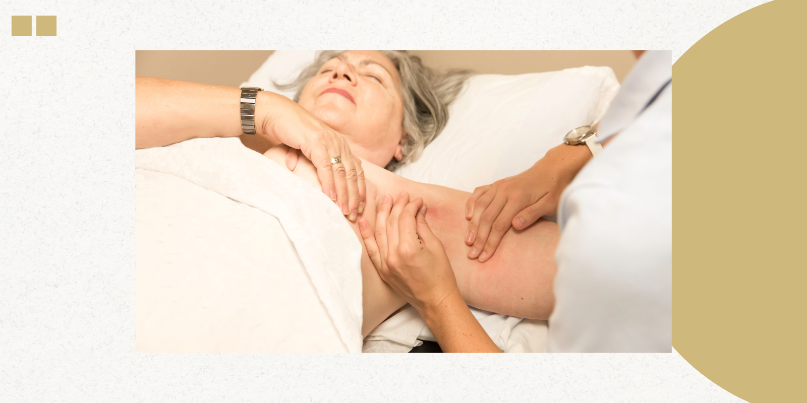
 Cytalux in action.
“When you give Cytalux as an infusion the day before or a few hours before surgery, it preferentially binds to the tumor and lights up the spot in the lungs,” Meguid says. “We use it largely in patients with metastatic cancers to their lung — for instance, colon cancer that has metastasized to the lung — because it helps us pick up spots that are too small to be seen with the naked eye.”
Not every patient is a good candidate for Cytalux, says Elizabeth A. David, MD, MAS, professor of cardiothoracic surgery, but for those with smaller tumors, the technology can be a boon for thoracic surgeons.
“Based on our recommendations for surgically treating lung cancers, when a tumor is less than two centimeters, the appropriate surgical treatment is doing either a segmentectomy — just removing a section of the lung — or a wedge resection, in which a small, triangle-shaped piece of tissue, including the tumor, is removed,” she says. “Those are the cases where we can use Cytalux to enable us to pinpoint where the tumor is and resect a minimal amount of lung tissue. We still do a lymph node dissection for those patients to make sure they're getting the appropriate operation.”
Going deep
A similar technology called Ion robotic navigational bronchoscopy uses a robotic bronchoscope to go deep into the lungs to find tumors, using fluorescent dye to identify them for biopsy or removal via robotic surgery.
 A surgeon uses ION during a procedure.
“We can get to spots in the periphery of the lung that we couldn’t get to in the past,” Meguid says. “The probe is about a millimeter thick, whereas a normal bronchoscope is about six millimeters thick. In the past, we were taking out larger sections of the lung, like a whole lobe, or part of a lobe. Now, we can take out smaller spots. If the spot is a precancer, we can stop at just that little wedge instead of larger section like we did in the past.”
Multidimensional view
Before surgeons even enter the operating room, a tool called Ceevra, which assembles CT scans into a 3-D image that can be rotated and zoomed in or out, allows them to better plan the exact route for a surgical robot to take.
“This 3-D reconstruction allows us to visualize the segmental anatomy of the lung to guide appropriateness of sublobar lung resections, which are becoming the main treatment for low-stage lung cancer,” Meguid says.
 David using Ceevra during an operation. The 3D image of the lung is on the laptop at right.
CT scans are taken six to eight weeks before surgery; the 3D model is created around 48 hours before the procedure. Surgeons can view the image on their phone or tablet, not only helping them to plan the surgery, but also to explain to patients exactly where a tumor is located and how it will be removed.
That’s especially important in Colorado, she says, where the elevation means less oxygen, making it vital to leave as much lung tissue as possible.
‘Early adopters’
CU lung surgeons have been using Ion navigational bronchoscopy for three years, Meguid says, and Cytalux and Ceevra for around 18 months. He has seen the benefit the tools have for surgeons as well as patients.
“Two days ago, the Ceevra imaging helped me do a more appropriate operation for a patient, saving more lung tissue,” he says. “And Cytalux is helping us find spots that we wouldn't have found with the naked eye, so it’s helping patients have a better cancer outcome. This is really cutting-edge technology, and we are early, early adopters.”