
<img height="1" width="1" style="display:none" src="https://www.facebook.com/tr?id=799546403794687&amp;ev=PageView&amp;noscript=1">

How a Protein Helps Determine Which Brain Cells Get Myelinated

Study reveals how gephyrin helps control myelin formation in the brain

minute read

by Chris Casey | October 16, 2026
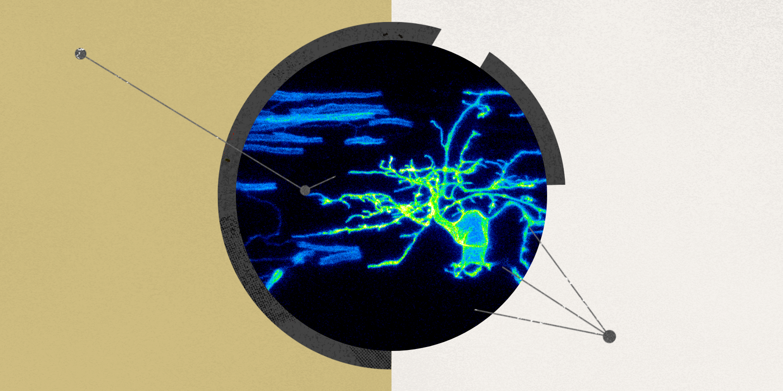
Stylized image of myelinating oligodendrocytes in living zebrafish larvae.
The takeaway:

Researchers at CU Anschutz have discovered that a protein called gephyrin helps determine which nerve fibers receive myelin, a protective coating that supports communication in the brain and spinal cord. The study, conducted in zebrafish, found that gephyrin influences where myelin forms and how long its sheaths grow. The findings offer new insight into how myelination may help regulate neural circuits, with potential implications for understanding neuropsychiatric disorders. 

Scientists have long known that disruptions in synaptic communication are a central part of the puzzle of neuropsychiatric disorders including autism, schizophrenia and bipolar disorder.

A research team led by Bruce Appel, PhD, professor of pediatrics-developmental biology at the CU Anschutz School of Medicine, wondered why people with these types of disorders often have abnormalities in myelin as well. Myelin is a protective, insulating coating around axons, the nerve fibers that carry signals in the brain and spinal cord.

The study published in Nature Communications showed how oligodendrocytes, which are glial cells in the central nervous system that produce myelin, help fine-tune neural circuits in zebrafish by controlling where myelin forms. “Myelination is not simply a hardwired process. There is a genetic regulation that provides a selectivity and specificity,” Appel said.

More specifically, the zebrafishes’ oligodendrocytes use the postsynaptic scaffold protein gephyrin to preferentially form myelin around inhibitory axons while simultaneously shortening the length of myelin sheaths they produce. 

What did the researchers discover about myelination?

  • Axon selection: Gephyrin helps oligodendrocytes preferentially myelinate inhibitory axons.
  • Sheath growth: Without gephyrin, myelin sheaths grow longer than normal.
  • Neural circuit balance: Changes in myelination may affect the balance between excitatory and inhibitory signaling, although the study did not directly test this effect. 

The study also found that in the absence of gephyrin, the oligodendrocytes myelinate the inhibitory axons less frequently and excitatory axons more frequently. Inhibitory and excitatory signaling work together to maintain balanced activity in the brain.

An excitation-inhibition imbalance is a well-researched neurobiological theory of autism spectrum disorder. “Our study raises the possibility that if you myelinate inhibitory axons less frequently and excitatory axons more frequently, that’s going to contribute to the hyper-excitation of the excitatory neural circuits,” Appel said.

Glossary:

Myelination – The process of forming myelin, an insulating coating around nerve fibers that helps electrical signals travel efficiently.

Oligodendrocytes – Specialized cells in the brain and spinal cord that produce myelin.

Gephyrin – A scaffolding protein that organizes other proteins involved in cell signaling and, as this study shows, helps regulate myelin formation.

Inhibitory axons – Nerve fibers from neurons that help reduce or restrain activity in neural circuits.

Excitatory axons – Nerve fibers from neurons that help increase activity in neural circuits.

GABAergic and glycinergic axons – Axons associated with neurons that use GABA or glycine, two inhibitory neurotransmitters.

Synapse – a junction where one neuron communicates with another cell.

The following Q&A further explains the study findings, including the suggestion that some of the molecular mechanisms involved in synapses may also help regulate myelin formation. The interview has been edited for length and clarity. 

Q&A Header

How does gephyrin help determine which axons receive myelin?

Selectivity comes from what we call cell adhesion molecules. It’s a molecule expressed on one partner and then a corresponding molecule expressed on another partner, and these can interact. It’s kind of like a zipper or Velcro. Gephyrin is not one of those molecules. It is on the inside of the cell membrane in the myelin sheath. We call it a scaffolding factor. It organizes the assembly of all these molecules that are important for synaptic activity. It assembles receptors that receive neurotransmitters (postsynaptic), the neural synapse and the cell-adhesion molecules and interacts with components within the cell. So gephyrin is like a hub, assembling all the other stuff that’s important. 

Why is it significant that some myelin sheaths contain large amounts of gephyrin while others contain little or none?

Yes, there’s more gephyrin in the myelin on the inhibitory axons than on the excitatory axons. Oligodendrocytes have many of these myelinating processes. In principle, gephyrin can go to any of them, and we think initially it is uniformly distributed, and then it becomes concentrated in the myelin on the inhibitory axons (GABAergic and glycinergic axons). We don’t know how that happens. One possibility is that it’s kind of like a capture mechanism. So these oligodendrocyte processes are extending, retracting and sampling their environments. So we think that maybe if it comes in contact with an inhibitory axon and it sees some kind of cue there – a cue to capture gephyrin there, which then begins to assemble all the other components. We’re years away from being able to test that because it’s going to require some heavy-duty engineering and microscopy.

But you’re right – there’s more gephyrin in the myelin, in inhibitory axons. If you take away the gephyrin, now these inhibitory axons don’t get myelinated as often, indicating that gephyrin really is important for that axon recognition. 

And if they aren’t myelinated as often, they don’t function as well?

That’s right – you have less inhibition. So the idea would be that with less myelin on them, they’re firing less frequently. Those neurons are less active, so you have less inhibition. And in the meantime, it’s not that the myelin doesn’t happen at all; it goes to the excitatory axons. So now those circuits are more active, and you’ve got more excitation. 

How does gephyrin regulate the length of myelin sheaths?

We actually think there’s two things going on. One is the axon specificity or the selection, and two is regulating sheath growth. In the absence of gephyrin, when the sheaths form, they get too long. We have other genes that we work with that, if taken away, the sheaths are too short. And here’s the interesting thing that we don’t understand: If you mutate the proteins that are implicated in the inhibitory synapses like gephyrin, those sheaths are consistently too long.

If you mutate the components that are associated with excitatory synapses, those sheaths are too short. So there’s some sort of balance between this machinery that’s determining myelin sheath growth, and we don’t understand this yet. We have some ideas for the excitatory side, but we haven’t tested it yet. That’s going to be a big push over the next few years. We think it’s interaction with all of these other receptors and signaling molecules that’s kind of controlling how that sheath grows.

So the idea is that you have elevated levels of neural activity, have more myelination on certain circuits. We haven’t gotten down to testing that really well yet. But the idea is that this machinery that responds to the neuronal activity can change the myelin sheath characteristics. You can shorten them and you can lengthen them to adjust the circuit activity.

And that’s exactly what happens at neuronal synapses. These same components respond – these molecular assemblies change to strengthen or weaken synaptic strength. We think this similar sort of thing is going on with myelination, but we haven’t tested it yet. 

How will researchers test gephyrin’s effects on myelination?

These proteins are genetically encoded, so we can make zebrafish lines. So we can silence or activate individual axons and then study their myelination because the real strength of our system is that through time-lapse imaging, we can watch these fluorescent proteins in specific cell types. And then we can manipulate those factors that we think are mediating (neurodevelopment mechanisms) and ask if those changes in myelin sheath characteristics and response to neural activity require these factors. 

What are the study’s limitations?

The big limitation of this study is that we weren’t able to test whether if you myelinate inhibitory axons less frequently and excitatory axons more frequently, you’ll end up with a hyper-excitation of the excitatory neural circuits. That’s because we need to be able to create zebrafish lines that eliminate gephyrin in all of the oligodendrocytes and only in the oligodendrocytes. 

What is the most important finding about how myelin forms?

That myelination is not simply a hardwired process, but one that’s nuanced: The molecular composition and placement of myelin may be tailored to particular neuronal classes, potentially allowing oligodendrocytes to participate in fine-tuning neural circuits. There is a genetic regulation that provides a selectivity and specificity (on the variable lengths and thicknesses of myelin on individual axons) that we hadn’t appreciated before.

Image at top: Myelinating oligodendrocytes in living zebrafish larvae. 

Key points:

- CU Anschutz researchers identified a role for gephyrin in controlling where myelin forms along nerve fibers.

- In zebrafish, gephyrin helps oligodendrocytes preferentially myelinate inhibitory axons.

- Gephyrin also helps regulate myelin sheath length, suggesting that myelination is more selective than previously understood.

- The findings raise questions about how myelination may influence the balance of excitatory and inhibitory activity in neural circuits.

- Further research is needed to determine whether these changes directly affect neural circuit activity and how they might relate to human neuropsychiatric disorders.

Featured Experts
Staff Mention

Bruce Appel, PhD