Using laboratory engineered tissue, scientists at the University of Colorado Anschutz Medical Campus have created a full thickness, biodegradable patch that holds the promise of correcting congenital heart defects in infants, limiting invasive surgeries and outlasting current patches.
The findings were published this week in the journal Materials Today.
“The ultimate goal is to make lab-grown heart tissue from a patient’s own cells that can be used to restructure the heart to correct for heart defects,” said the study’s senior author Jeffrey Jacot, PhD, associate professor of bioengineering at the University of Colorado School of Medicine.
About 10,000 babies are born with a complex congenital heart defect every year in this country, requiring surgery in the first year of life. Some of these operations require the implantation of a full-thickness heart patch. But the current materials used in the patch are non-living and non-degradable. They don’t grow with the patient and often fail because they don’t integrate with the heart.
Jacot said these surgeries are largely palliative, extending survival only until the next surgery.
New patch engineered for longevity
But his lab’s patch, called a tissue engineered myocardial patch, could survive the mechanical forces of the heart wall and integrate into the heart itself. Ideally, it would last for as long as the patient lives.
“The current patch materials available to pediatric heart surgeons are exclusively non-living and non-degradable, which often fail in their long-term therapeutic efficacy due to low compliance, an increased risk of thrombosis and intimal hyperplasia, and their inability to remodel and integrate with the heart,” the study said.
Permanent fixes require biomaterials that are degradable but that also promote heart regeneration so that the patches are eventually replaced by healthy myocardium, the middle muscular layer of the heart and the thickest.
“Any patches that are not replaced by healthy tissue prior to their degradation will inevitably fail and lead to long-term complications,” Jacot said.
More testing needed before human trials
The patch was created in the lab using a technique known as electrospinning, where electricity is applied to liquid solutions to create nanofibers used to make a "scaffold." The scaffold is then injected with living cells. This eventually becomes the patch.
“The scaffold was found to be mechanically sufficient for heart wall repair,” Jacot said. “Vascular cells were able to infiltrate more than halfway through the scaffold in static culture within three weeks.”
The patch requires more tests before it can be used in humans.
Jacot is optimistic that it will play a critical role in the future treatment of congenital heart defects and other cardiac conditions.
“This is the first successful demonstration of a very thick, porous electrospun patch specifically for cardiac tissue engineering,” he said.
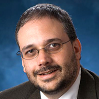
Photo at top: Jeffrey Jacot, PhD, poses in his lab.


