.png)
When a patient with lymphoma lost vision in one eye, neuro-ophthalmologist and orbital surgeon Prem Subramanian, MD, PhD, was concerned that it was because the cancer had infiltrated the optic nerve, the cord connecting the brain and eyes. Yet, every test — blood tests, MRI scans, and a spinal tap — delivered inconclusive results on whether that was the case. Then, the patient began losing vision in their other eye. Time was of the essence to save what sight the patient still had, leading Subramanian to make the tough decision to perform a high-risk procedure.
“I did a full-thickness biopsy of the optic nerve, removing a piece of the optic nerve from the side that was connected to the already blind eye,” says Subramanian, professor and chief of neuro-ophthalmology at the University of Colorado Anschutz Department of Ophthalmology. “The biopsy gave us the answer that lymphoma was affecting the patient’s optic nerve, and that allowed the patient to move forward with treatment and preserve their remaining vision.”
An optic nerve biopsy is often a last resort because of the significant risks it poses, raising the question of how valuable these biopsies are. Research published in 2025 found that out of a total of 72 patients who received an optic nerve biopsy or optic nerve sheath biopsy at a Mayo Clinic location, 67% of them received a diagnosis. Hoping to raise awareness of this research, Subramanian, who helped review the study before it was published, wrote an editorial piece published in the Journal of Neuro-Ophthalmology, “Optic Nerve Biopsy — Is It Ever Useful?”
“It may seem like a procedure you’d never want to do because of the risk, but I think these biopsies are useful,” says Subramanian, who is also the Clifford R. and Janice N. Merrill Endowed Chair in Ophthalmology. “I have done optic nerve and nerve sheath biopsies throughout my career, and so it was very important to me that this research be shared with other doctors so they can better understand how to do this, when to do it, and what kind of information they can gather from it.”
The eye-brain connection
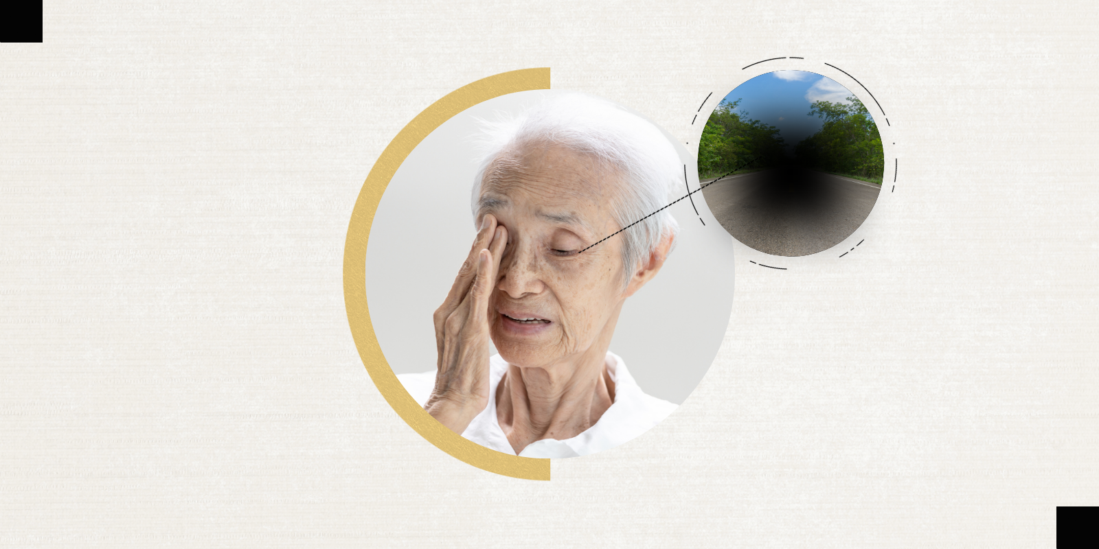
The optic nerve is a fragile but critical component of vision. There are more than 1.1 million retinal ganglion cells (a type of neuron) that exist in the retina, which is a layer of tissue near the back of the eyeball that detects light. Each retinal ganglion cell has a long axon that connects to the brain, similar to a long cable. The axons of retinal ganglion cells combine to form the optic nerve. These neurons take information captured by the retina and send it to the brain so it can process and understand what the eyes are seeing, such as movement and color.
Damage to the optic nerve is often considered permanent, underscoring the value of neuro-ophthalmologists like Subramanian.
“My specific interests are optic nerve disorders, such as inflammation, infection, blood supply or vascular problems, and tumors that compress the optic nerve,” he says. “There is also a significant percentage of patients who have tumors that cross between the orbit and the intracranial space. Those tumors can affect eye movement or vision, so I will often collaborate with neurosurgeons and ear, nose, and throat specialists to provide surgical and medical care.”
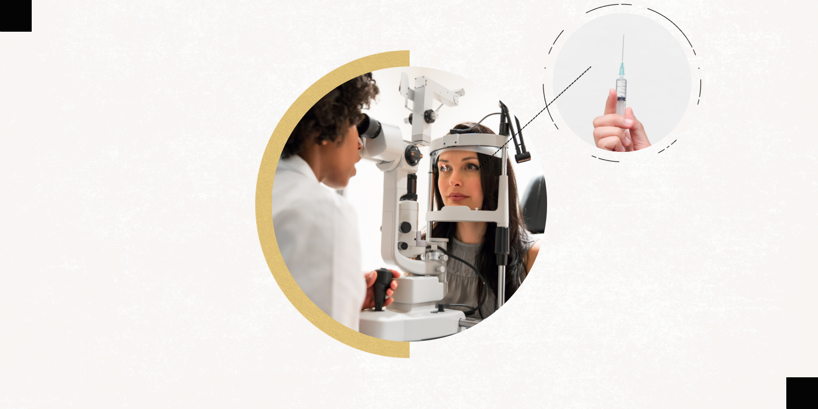
However, sometimes the issues affecting the optic nerve are unclear. It’s in these cases that a biopsy of the optic nerve or the optic nerve sheath may be considered.
“Because these biopsies carry a high risk of damaging a person’s vision, it’s typically done in people where every other way of making a diagnosis has been tried, and we still don’t have an answer as to why they have a vision problem, but we know that it’s an issue with the optic nerve,” he says.
Deciding what to cut
An optic nerve sheath biopsy is generally less invasive and risky compared to a biopsy of the optic nerve itself. The structure of the optic nerve is essentially like a cable, and the nerve sheath is somewhat similar to insulation surrounding the cable, almost like a cover, Subramanian explains.
The sheath is normally very thin, but it sometimes can become thicker if there is a disease affecting it. It is possible to do a biopsy of the sheath without affecting the optic nerve itself.
“Deciding how to do a biopsy is critical,” Subramanian says. “It’s important to consider whether the disease is likely to be affecting the sheath, the optic nerve, or both.”
For the optic nerve, there is a difference between a partial and full-thickness biopsy. A full-thickness biopsy means that the surgeon cuts all the way through the optic nerve, a procedure that effectively blinds the eye connected to that cord. This is a more viable option for patients who are already blind in one eye. Since there is an optic nerve behind each eye, it’s possible to do a full-thickness biopsy of the blind eye’s optic nerve without damaging the other eye that can still see.
“In other cases where the patient still has vision and you can’t do a full-thickness biopsy, you have to balance out the likelihood of getting an answer and the preservation of vision,” Subramanian says, explaining that a common error surgeons make is taking too small of a sample.
“The research showed us that if you do a timid biopsy and only remove a small amount of tissue, then you probably shouldn’t even do the biopsy at all because you’re unlikely to get a diagnosis from it,” he says. “You have to take an adequate specimen in order to make the risk of the biopsy worth it to the patient.”
Overall, the Mayo Clinic research found that optic nerve biopsies were more likely to result in a diagnosis compared to nerve sheath biopsies (81% compared to 55%). The findings also suggest that full-thickness biopsies are more likely to uncover answers than partial thickness or lower tissue biopsies.
“The procedure itself can be done in a number of different ways,” Subramanian says. “We use different orbital surgery approaches depending on whether the area of interest is right behind the eye, in the middle of the optic nerve, or at the back of the orbit. Sometimes, we work with neurosurgeons to operate on the part of the head where the optic nerve is starting to enter the intracranial space. It really depends.”
Protecting patients’ sight
In his 25-year career, Subramanian estimates that he has done these biopsies less than 10 times. Though rare, these procedures can help patients in big ways, he explains.
One of his patients, for instance, was experiencing progressive vision loss and it was unclear why, with the main suspicions being inflammation or a potential tumor. After numerous tests and a failed steroid treatment, Subramanian conducted an optic nerve sheath biopsy that confirmed the patient had a tumor, allowing her to move forward with getting the appropriate treatment.
These biopsies, however, do not always result in a clear diagnosis. For example, Subramanian had another patient who went blind in one eye and the other eye was losing vision. He performed an optic nerve biopsy on the patient’s blind eye that showed non-specific damage to the optic nerve, meaning the underlying cause of the damage was still unclear.
“But that biopsy was still helpful because it allowed us to confirm there was not a malignant tumor within the optic nerve — it excluded that concern,” he says.
In future research, Subramanian hopes to see further genetics studies that help neuro-ophthalmologists develop better pathology techniques so they can make more diagnoses from these biopsies.
“Overall, these biopsies shouldn’t be done if you can get answers another way,” he says. “But for the right patient, it does make sense to do this because it can help us preserve and protect the patient’s sight.”