.png)
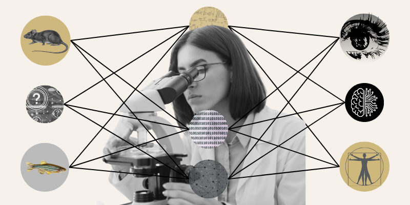
What if we could read the hidden language of cells? Cell Painting is making that possible. This cutting-edge technique is helping researchers uncover secrets hidden inside our cells. Recently, Jenna Tomkinson, a biomedical informatics researcher from the Way Lab at the University of Colorado Anschutz (CU Anschutz) School of Medicine, brought this colorful science to life during a presentation at National Jewish Health, where she introduced the audience to the world of high-content imaging and its role in drug discovery.
What Is Cell Painting?
Using fluorescence microscopy, scientists “paint” cells with different dyes to highlight their structures. These images aren’t just pretty pictures; they’re packed with data. By analyzing these images, researchers can detect subtle differences between cells that the human eye would miss. This approach is revolutionizing phenotypic profiling and helping scientists predict how cells respond to drugs.
For Tomkinson, the appeal of this work is its ability to uncover what the eye can’t see. “Over time, I’ve learned that this methodology is really robust and can discover the little nuances between cells that we can’t always see with our eyes,” she explained. “I find this field to be very innovative, and I hope to contribute more as I progress in my role.”
Why It Matters
The Way Lab’s work doesn’t stop at pretty images. Their research is about accelerating the search for new treatments. By turning cell images into measurable data, researchers can identify promising compounds long before they reach clinical trials. This early-stage work is critical because it helps narrow down thousands of possibilities to the few that might truly make a difference for patients.
Tomkinson views this as a way to bridge the gap between discovery and care. “Our work serves upstream from patient care,” she explained. “Our impact comes slower than does in clinical trials, but the work we do sets up our clinical colleagues as we work together to identify treatments for disease.”
Building Connections Through Science
During her session, Tomkinson walked attendees through the fundamentals of Cell Painting and shared insights from a recent study on NF1 genotypes in Schwann cells. Using machine learning, her team transformed cell images into data points, uncovering biomarkers that could guide treatment strategies.
The presentation sparked lively discussion. When asked if biologists could easily adopt these techniques, Tomkinson shared, “Not easily, but it can be done. When I started my role three years ago, I had no idea how to do everything I do today, but I was able to learn quickly by using the resources around me.”
.png?width=800&height=400&name=Newsroom%20Header%20(11).png)
This workshop wasn’t just about sharing science; it was about building connections. National Jewish Health and CU Anschutz have a long history of collaboration, and this event paved the way for new partnerships. Tomkinson described the experience as a privilege: “I got to be the expert on image analysis and image-based profiling and teach other researchers about why our work is important and could be something that they can utilize in their work.”
Several researchers expressed interest in applying these techniques to their own projects, especially in tissue research. As Tomkinson noted, “All it takes is an email to spark a collaboration.”
Stay connected by following the Way Lab on LinkedIn, visiting their website, or exploring open-source work on GitHub.