When a young child comes to a hospital emergency department with serious head trauma, medical providers must make a quick decision that could have major consequences: What’s the best test to find out if they have a brain injury?
That’s the question that a University of Colorado Anschutz Department of Emergency Medicine trainee and a faculty mentor address in a new study published in the American Journal of Neuroradiology.
The lead author is Liam Canty, MD, who was a CU Anschutz School of Medicine student while working on the study and is now a first-year emergency medicine resident. The senior author is Daniel Lindberg, MD, a professor of emergency medicine and a member of the department’s Family Violence Recognition Lab.
In particular, Canty and Lindberg wanted to assess whether a “fast MRI” – a shortened version of a standard magnetic resonance imaging scan that takes about six or seven minutes instead of half an hour or longer – can be an acceptable initial imaging method for small children with head trauma.
Fast versus routine MRIs
Both fast MRIs and full, “routine” MRIs use powerful magnets and radio waves to create detailed images of the body’s interior. Both use the same equipment but different protocols. Importantly, fast MRI avoids the need for anesthesia, which is often required to keep children from moving during longer, routine MRIs.
In most emergency departments, the imaging used for these cases is a computed tomography (CT) scan, which uses X-rays and exposes children to radiation. An MRI doesn’t involve radiation.
“It’s been shown that sedative medications in very young children often required with MRIs can cause neurocognitive harms down the line, and there’s the radiation exposure with CTs, so obviously you’re trying to balance that,” Canty says.
“If you give a kid a bottle, wrap them up, and give them earplugs, they won’t move for a few minutes,” Lindberg says regarding fast MRIs. “That’s less true for 35 minutes.”
But what isn’t well established is whether a fast MRI will miss important injury findings that a routine MRI will catch.
“The idea that you might miss a brain injury in a kid is something that's very worrisome,” Lindberg says.
A good mentor
According to the Brain Injury Association of America, about 435,000 children age 14 and younger are seen in emergency departments each year with suspected brain injuries, leading to 37,000 hospitalizations and an estimated 2,685 deaths. Research shows that children under age 5 face the highest rate of ED visits for head injuries among age groups, usually due to falls. Abusive head trauma, formerly called “shaken-baby syndrome,” is a leading cause of child abuse deaths in children under 5.
→ Daniel Lindberg on Using AI to Find Red Flags for Physical Abuse of Children
The fast MRI study grew out of discussions between Lindberg and Nicholas Stence, MD, a professor of radiology and a pediatric neuroradiologist who has been instrumental in developing fast MRI techniques. Stence was a co-author of the new study and collaborated with Lindberg on earlier research on fast MRI.
While a med student at CU Anschutz, Canty sought out research opportunities and got in touch with Lindberg, who asked if he wanted to work on the fast MRI project.
“I was excited to get involved with it,” Canty says. “I wanted someone who was a good mentor to work with, and that’s how it started.”
Comparing MRI results
The study looked at data on 2,540 children age 6 and younger who came to a pediatric ED between 2014 and 2024 and had a head injury evaluation. Of that group, 69 got both a fast MRI and a full routine MRI within a week of each other.
That paired data allowed researchers to evaluate what both scans found in the same child. Imaging results from both types of MRI were compared across several categories, including various types of bleeding around the brain as well as injury to brain tissue.
The remaining 2,471 children in the study initially received only a fast MRI that turned up negative results for brain injury. Of that total, 1,122 came back to the ED or an inpatient clinic within one year for various reasons. The researchers checked if any of those children returned with a brain injury that the initial fast MRI missed.
Among the study’s key findings regarding the group of 69 children who were scanned twice:
- The fast MRI successfully detected at least one or more trauma-related abnormality in 68 children – a finding that led to the children being re-scanned in a routine MRI.
- Fast MRIs were found to be as good as a full MRI in detecting epidural hemorrhage (bleeding between the skull and the brain's outer lining), and did well for subdural hemorrhage (bleeding just under the outer lining).
- The fast MRI was not quite as sensitive as the routine MRI in detecting subarachnoid hemorrhage (SAH), a different kind of bleeding. The fast scans caught SAH in 75% of the 24 cases where a routine MRI later found it to be present.
- The fast MRI was also less effective in detecting damage to the brain itself – a parenchymal injury. It found such an injury in 64.5% of the 31 cases where the routine MRI picked it up. As with subarachnoid hemorrhages, these findings may have evolved and changed in the time between the two scans.
A reliable screening tool
Of the 1,122 children who returned to the ED or a clinic after a clear fast MRI scan, the study says only four were found to have a brain injury on follow-up imaging, and all of those cases were attributed to new trauma since the initial scan rather than something missed the first time around.
“We were trying to find out if we could have gotten a fast MRI that was negative, and they went home with a brain injury that got worse, and they had to come back,” Lindberg says. “We feel pretty comfortable that that wasn't the case.”
The study’s bottom line, Canty and Lindberg say, is that a fast MRI appears to be a reliable initial screening tool that’s unlikely to entirely miss indications of brain injury, even if it isn’t as thorough as a routine MRI in picking up more subtle findings.
“There was never an isolated parenchymal injury the fast MRI missed that wasn't also accompanied by another injury,” Canty says. “Overall, it has a very high sensitivity for picking up a head injury, if maybe not as great of specificity when compared to routine MRI.”
Fast MRI, they say, can spare many children from unnecessary radiation or sedation while still catching the injuries that matter most.
“This was almost 2,500 kids who weren’t exposed to radiation, and that’s maybe a case or two of cancer prevented,” Lindberg says.
Fast MRI as an initial test
Canty and Lindberg suggest that clinicians consider using fast MRI as an initial screening test, and if the results come back positive or unclear, or if child abuse or a skull fracture are suspected, providers should follow up with a routine MRI or a CT scan for a complete picture.
Lindberg says that in some cases, it would still be best to proceed directly to a routine MRI or CT scan. “If a kid's in a coma, or if they're really unstable, I'm not going to do a fast MRI on that kid first. They've got bigger risks to worry about than the radiation or the sedation.”
“We would love to see fast MRI become more common,” Canty says. “Obviously, a hospital has to have the resources necessary to do a fast MRI. Coming from a rural area in Colorado myself, I know this is something that's not always practical. But in the places where it’s available, we would love to see it.”
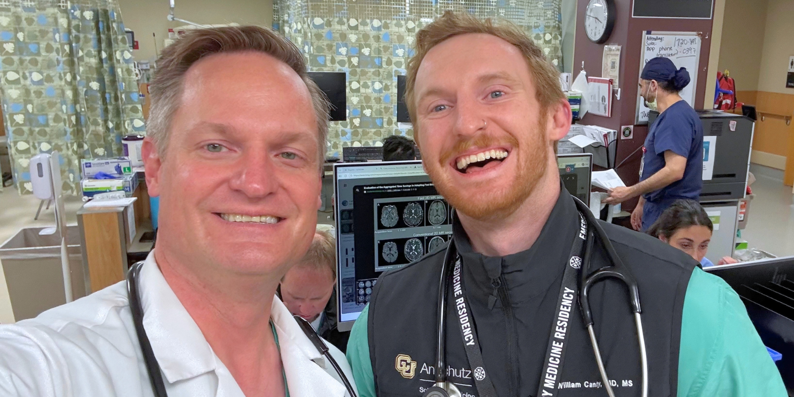
Photo at top: Daniel Lindberg, MD, and Liam Canty, MD.