Three University of Colorado Cancer Center scientists have received a combined total of almost $2 million in grant funding from the American Cancer Society (ACS) to support research addressing a broad spectrum of cancer prevention, diagnosis, and treatment.
The ACS is the largest private, not-for-profit source of funding for scientists studying cancer in the United States, funding basic, translational, clinical, and cancer control research. Support from the ACS aids CU Cancer Center scientists in remaining at the forefront of discovery and innovation in cancer research.
Risk of pediatric AML relapse
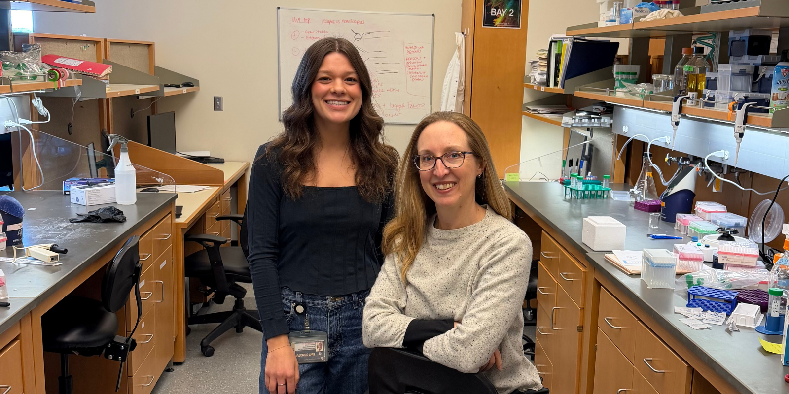
Among the research receiving ACS support is a study seeking to improve clinicians’ ability to determine pediatric acute myeloid leukemia (AML) patients’ risk of relapse early in treatment. Amanda Winters, MD, PhD, assistant professor of pediatric hematology, oncology, and bone marrow transplantation in the CU School of Medicine, will work to develop tests that can measure genetic measurable residual disease (MRD) in these patients, with a goal of earlier intervention for patients at high risk of relapse.
“One child is diagnosed with AML every day in the United States,” Winters explains. “Despite intense chemotherapy and bone marrow transplant, about one-third of these children will ultimately relapse with difficult-to-treat disease. Improvement in doctors’ ability to determine an individual patient’s risk of relapse early in treatment can assist in tailoring therapy to best treat that patient’s disease and can identify those who may benefit from increased or decreased intensity of therapy.”
Winters and her co-researchers will work to develop three different tests for genetic MRD, which has been proven to be the best predictor of relapse in patients receiving therapy for AML. Currently, the most commonly used test for MRD is flow cytometry, which identifies abnormal protein expression on leukemia cells compared to normal cells.
“The ultimate goal of this project is to provide additional clinical tools for doctors to risk stratify patients so that therapy can be designed to give each patient the best chance of long-term survival without relapse,” Winters says.
Developing an mHealth cancer prevention tool
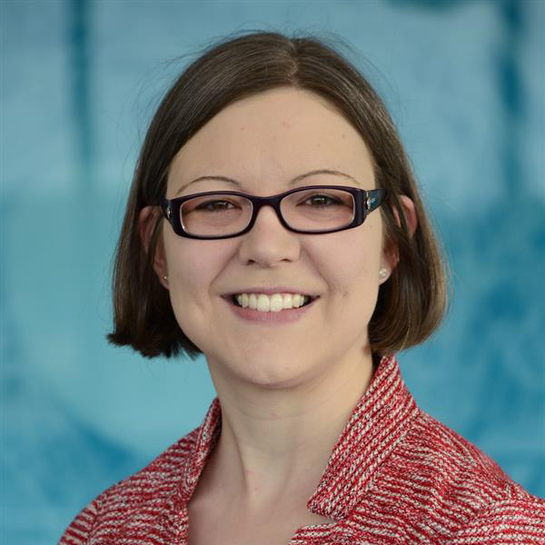
Swati Patel, MD, MS, associate professor of gastroenterology in the CU School of Medicine, will lead research studying the yield of expanding genetic evaluation to people with advanced polyps, which are the immediate precursor to colorectal cancer. A further research goal will then be to design a mobile and digital health (mHealth) intervention that empowers patients to orchestrate their prevention care.
“Based on our preliminary data, we expect that similar to colorectal cancer, 10% of advanced polyp patients will be diagnosed with a hereditary syndrome,” Patel says. “Hereditary cancer syndromes are a common cause of multi-organ cancers. Although there are effective cancer prevention strategies, these syndromes are grossly under-diagnosed and patients with these syndromes feel overwhelmed by complex care programs, leading to missed opportunities for cancer prevention.”
Patel and her co-researchers will recruit advanced polyp patients to complete genetic testing, then conduct detailed interviews to explore the spectrum desired mHealth content, platform, and delivery options to promote using cancer prevention care.
“This study will provide the data needed to expand genetics services to those with advanced polyps and produce an mHealth intervention to improve multi-cancer prevention care among patients with hereditary syndromes,” Patel explains.
Using cell-free DNA to benefit immunotherapy
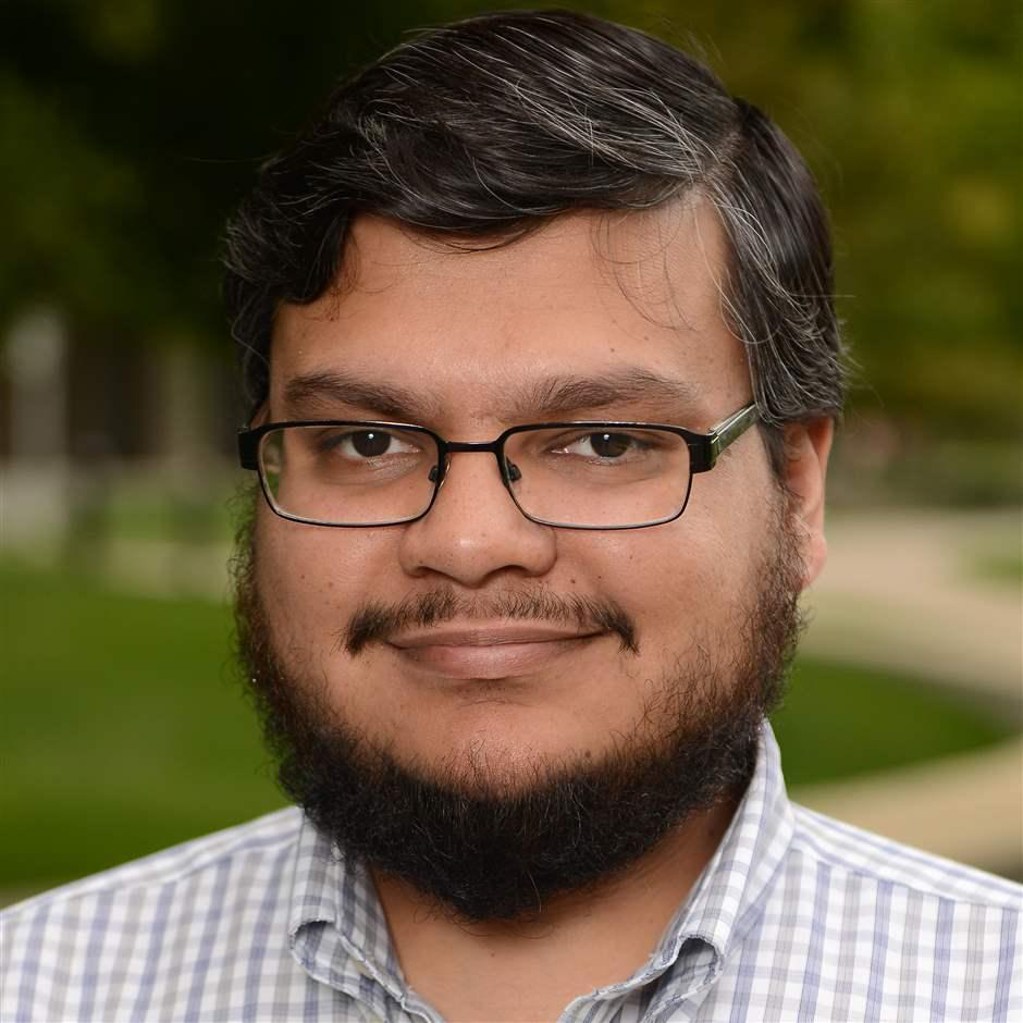
Researcher Srinivas Ramachandran, PhD, assistant professor of biochemistry and molecular genetics in the CU School of Medicine, is working to predict and track response to immunotherapy treatments by developing a more sensitive and less expensive method for generating gene expression signatures for cells that shed cell-free DNA (cfDNA).
“Cell-free DNA arises from genomic DNA shed by cells during turnover, which then ends up in blood, saliva, and urine,” Ramachandran explains. “Tumor cells, in particular, shed a lot of DNA, so harnessing information from cfDNA to identify which cells are shedding their DNA could be used to track cancer.”
Normally, cfDNA is generated during normal blood cell turnover, a process that dilutes DNA from a tumor when one is present. The human genome is packaged with proteins called histones, which form discrete units called nucleosomes. cfDNA is a snapshot of where nucleosomes were arranged on a dying cell’s genome.
Ramachandran and his co-researchers will use the length of cfDNA fragments near the start of genes to infer whether the gene was expressed in the cell that shed the cfDNA in the first place. Knowing which genes are on and off will enable them to predict the cell type based on known gene expression datasets.
By generating gene expression signatures for cells that shed cfDNA, Ramachandran’s goal is to better identify patients who will benefit from immunotherapy and, once it’s started, identify earlier whether the treatment is working.


